

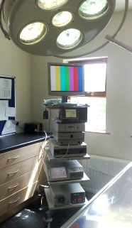
Specialist Veterinary Arthroscopy equipment
Stryker 1088 3 chip camera and console
Dynonics 1.9mm, 2.4mm & 2.7mm arthroscopes
Stryker xenon light source
Styker TPS & Shaver
Arthrocare bipolar / RF coblation
Arthrex Continuous Wave III arthroscopy Pump
Stryker SDC 2 image recorder
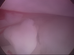
Elbow Arthroscopy- fragmented coronoid process FCP

Because arthroscopy is a minimally invasive surgery, both elbows can be examined and treated arthroscopically at one surgery. Due to the small surgical sites of 3-4 mm diameter there is greatly reduced post operative pain compared to open joint surgery so most dogs are walking on the operated legs immediately after surgery, and recover more quickly that after open surgery.
Effective pain medications are given intra-articularly immediately post op and followed up with injectable and oral medications for several days
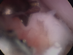
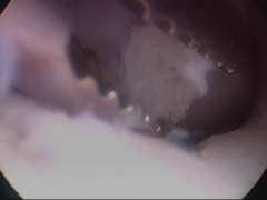
Arthroscopic examination of the elbow of a 2 year old Rotweiller dog with foreleg lameness. Multiple fragments of the coronoid process are removed followed by arthroscopic subtotal coronoidectomy (removal of part of the coronoid bone).
These loose fragmented coronoid process lesions which move on weight bearing, are a cause of persistent pain & lameness. Both the fragmented cartilage lesions and the underlying yellowish coloured avascular subchondral bone will be removed arthroscopically.The yellowish coloured avascular bone can be seen on the underside of the white cartilage covered fragments and its exposure within the joint will lead to significant DJD. Avascular bone is bone which has lost it blood supply.
The surgical instruments used arthroscopically are very small- approx 2-3 mm in size and the detail of magnification is seen in the videos- during the surgery this view is displayed in HD resolution on a 19” stryker monitor to allow accurate diagnosis and surgery.
Arthroscopic surgery allows detailed visualisation and surgical treatment of lesions which cannot be seen clearly at open joint surgery with the benefit of less post operative pain, faster recover and very small 3mm operative sites.
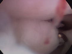
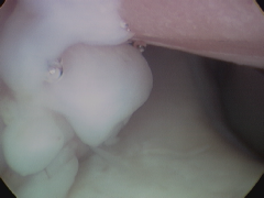
Once the fragmented coronoid lesion on the ulnar bone has been removed, the view indicates the region where the lesion had been sitting in the foreground and in the background we can see the normal cartilage covering the radial head- which is moving as we rotated the leg.The edges of the lesion will now be freshened to leave a smooth edge using either a hand burr or a motorised shaver and then a partial removal of the remaining coronoid process in the region of the lesion is performed using a motorised shaver (subtotal coronoid ostectomy SCO), and the joint is then thoroughly flushed to remove any loose cartilage fragments.
In cases of FCP with significant cartilage lesions affecting the humerus bone (kissing lesions) or in cases of co-existing OCD of the medial humeral condyle a proximal ulnar osteotomy may also be recommended in addition to fragment removal and subtotal coronoidectomy. Ulnar osteotomy involves cutting the ulnar bone at an oblique angle below the elbow joint and allowing it to heal over the following weeks. This additional treatment results in significant short term lameness while the ulnar heals but may benefit the dog long-term by allowing an improvement in joint incongruity.

Various treatments are recommended for FCP and veterinary opinions vary and alternative methods of treatment are currently undergoing clinical trials. Unfortunately the exposure of the bone underneath the cartilage (subchondral bone) to the joint, will stimulated the development of osteoarthritis. No treatment is available which will prevent osteoarthritis developing once it has begun, and all cases will require management of the elbow osteoarthritis by weight control, exercise modulation (regular controlled exercise), NSAIDs (anti inflammatory) medications and pain control.
| elbow arthroscopy key hole surgery dog ireland |
| elbow dysplasia |
| vet-arthroscopy-shoulder ocd-dog |
| shoulder arthroscopy dog- ligament injury |
| stifle arthroscopy dogs |
| hip arthroscopy dog |
| spinal xray myelogram |
| GSDA A stamp |
| elbow score |
| hip score |
| hip score guide for owners |
| directions |
| site map |